Muscle Tear While Playing Sports? How an Ultrasound Scan Can Diagnose and Guide Recovery
Experiencing a sudden snap or pull during a football match, gym session, or sprint can be alarming. It often signals a muscle tear, a common yet painful sports injury that can range from a mild strain to a complete rupture. Prompt and accurate diagnosis is key to ensuring proper healing—and that's where ultrasound imaging becomes a game changer.
What Is a Muscle Tear?
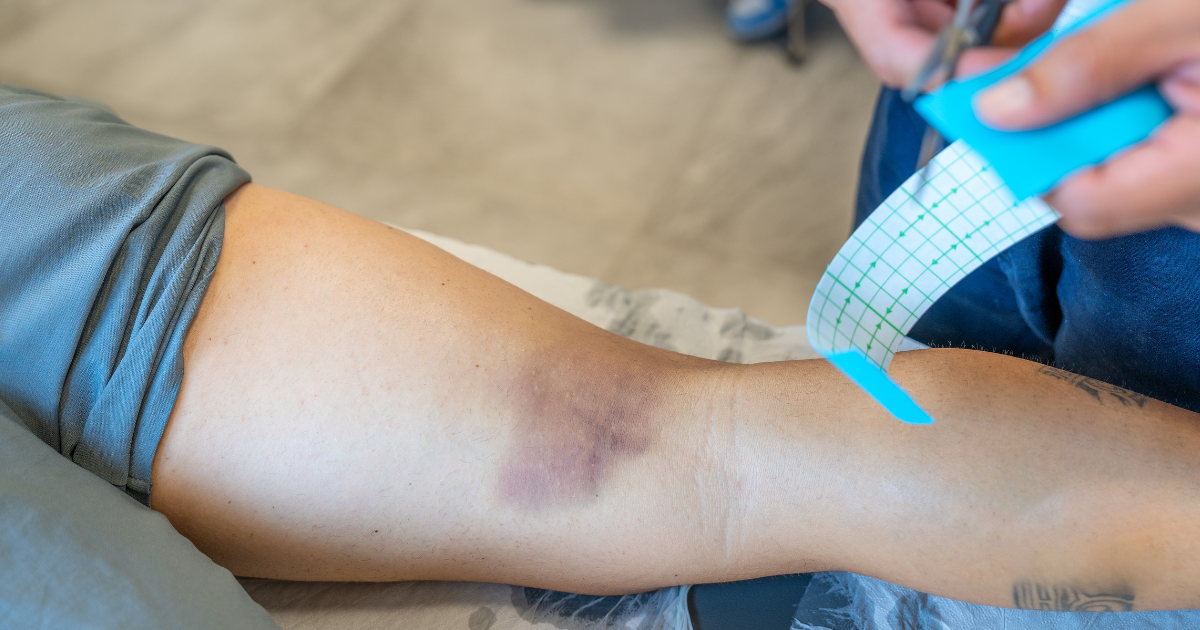
A muscle tear, also known as a muscle strain, occurs when the fibers of a muscle are overstretched or torn. This typically happens during sudden, forceful movements—especially in high-intensity sports like football, rugby, or sprinting. The affected muscle may become painful, swollen, weak, or bruised, depending on the severity of the injury.
Grades of Muscle Tear
Muscle tears are classified into three grades based on their severity:
Grade I – Mild
A small number of fibers are stretched or torn.
Minimal pain and swelling.
No significant loss of strength or function.
Grade II – Moderate
A larger number of muscle fibers are torn.
Noticeable pain, swelling, and bruising.
Limited movement and strength.
Grade III – Severe
Complete rupture of the muscle or tendon.
Severe pain, possible deformity (e.g., a lump or gap in the muscle).
Major loss of function; often requires surgery.
How Ultrasound Identifies the Grade of a Muscle Tear?
Ultrasound is a powerful diagnostic tool for grading muscle injuries based on the extent of structural damage:
Grade I (Mild): Ultrasound may show localized swelling or slight disruption of muscle fibers, often with hypoechoic (dark) areas indicating edema. There is no visible tear, and muscle continuity is preserved.
Grade II (Moderate): A partial tear is visible on ultrasound as a clear discontinuity in muscle fibers, with possible hematoma formation. The area may appear more hypoechoic due to fluid collection and fiber disruption.
Grade III (Severe): A complete tear shows as a full rupture of the muscle with retraction of the muscle ends. A hypoechoic or anechoic gap is visible between the torn fibers, and hematoma or fluid accumulation is often present.
Dynamic ultrasound (performed during muscle contraction or stretching) can further improve the accuracy of grading by visualizing functional impairment in real time.
Why Choose an Ultrasound Scan?
Traditional imaging methods like X-rays can’t visualize soft tissues, but musculoskeletal ultrasound excels in capturing muscles, tendons, and ligaments in real time. Here’s why it’s ideal for athletes:
1. Real-Time, Dynamic Imaging
Ultrasound allows clinicians to observe the injury during movement. For example, flexing a knee or rotating a shoulder can reveal hidden tears or instability that static scans like MRIs might miss . This dynamic assessment is invaluable for diagnosing conditions like hamstring strains or rotator cuff tears .
2. Non-Invasive and Radiation-Free
Unlike CT scans or X-rays, ultrasound uses sound waves, making it safe for repeated use—even for pregnant athletes or children.
3. Pinpoint Accuracy
Ultrasound provides high-resolution images, distinguishing between partial and complete tears. For instance, it can detect a small Achilles tendon tear or a subtle muscle hematoma with precision .
4. Guided Interventions
Beyond diagnosis, ultrasound directs treatments like corticosteroid injections, platelet-rich plasma (PRP) therapy, or fluid aspiration. This ensures medications reach the exact injury site, improving outcomes.
When Should You Get an Ultrasound?
Consider an ultrasound scan if you:
Heard or felt a “snap” during activity
Experience swelling, bruising, or difficulty moving the muscle
Can’t bear weight or use the limb normally
Notice a visible deformity or lump in the muscle
Early imaging leads to early action—essential in returning to sports safely.
Evidence-Based Insight
A 2021 review in Ultrasound in Medicine and Biology confirmed that ultrasound is a highly sensitive tool for evaluating muscle injuries, particularly in the acute and subacute phases. The review emphasized ultrasound's ability to detect even small tears early in the injury process, making it essential for timely diagnosis and treatment. Additionally, ultrasound's real-time imaging capability allows for monitoring tissue healing and remodeling, which is invaluable in sports rehabilitation. This continuous monitoring helps track recovery progress, assess scar tissue formation, and guide targeted interventions such as injections, making ultrasound an essential tool for ensuring safe and effective recovery.
How Ultrasound Guides Recovery
1. Tailored Treatment Plans
PRP and Stem Cell Therapy: Ultrasound guides injections of regenerative agents into the tear, accelerating healing. Studies show PRP reduces recovery time in athletes with muscle injuries.
Aspiration of Hematomas: Chronic blood collections in muscles (e.g., tennis leg) can be safely drained under ultrasound guidance, relieving pain and preventing scar tissue.
2. Monitoring Healing Progress
Regular ultrasound scans track tissue repair. For example, reduced swelling and improved fiber alignment indicate successful rehabilitation.
3. Rehabilitation Support
Physical therapists use ultrasound to design movement-based rehab programs. Real-time feedback ensures exercises don’t strain healing tissues.
Conclusion:
Ultrasound imaging is a highly effective and non-invasive tool for diagnosing and managing muscle tears, particularly in sports-related injuries. By providing real-time, dynamic imaging, ultrasound enhances the accuracy of diagnosis and allows for better monitoring of muscle recovery. Its ability to detect even small tears early and track tissue healing over time makes it invaluable in sports rehabilitation.
While ultrasound alone may not cure the injury, it plays a vital role in guiding treatment decisions, such as injections or physical therapy interventions, to accelerate recovery. A tailored approach based on the severity of the muscle tear and the patient's specific needs ensures the most effective outcomes.
When used properly, ultrasound can significantly reduce recovery time and help athletes return to their sport safely, ultimately improving quality of life and preventing long-term complications.
References:
Cummings, T. M., & White, A. R. (2015). The effectiveness of trigger point injections for myofascial pain syndrome: A systematic review. Clinical Journal of Pain, 31(10), 906-914.
D'Addona, A., Gabriele, A., De Bartolomeo, G., Parisi, A., & Manzoli, L. (2021). Ultrasound imaging in the assessment of muscle injuries: A systematic review and meta-analysis. Ultrasound in Medicine & Biology, 47(3), 673-682.
Smeets, R. J., & Baert, A. L. (2020). Ultrasound-guided injection therapy for musculoskeletal pain: A systematic review of clinical effectiveness and safety. European Journal of Pain, 24(10), 1959-1968.
Bhatia, R., & Wright, G. R. (2022). The role of musculoskeletal ultrasound in sports injury management: Current trends and future directions. British Journal of Sports Medicine, 56(6), 331-338.
Connell, D. A., & Koulouris, G. (2020). Musculoskeletal ultrasound: An overview of its role in diagnosis and management of soft tissue injuries. American Journal of Roentgenology, 214(3), 610-617.


